 Fuhua Chen
Analysis of tissue volumes in human brains, and its relation to some diseases based on magnetic-resonance-imaging (MRI) image processing
It has been widely recognized that by tracing and analyzing the change of the volumes of different tissues in human brains, some brain-related diseases can be found earlier so that some measure can be taken to stop the disease at its early stage. For example, it’s found that Alzheimer, a kind of intelligence disease for senior people, is currently very hard to cure. In many cases, doctors can only stop or slow the development of the disease. Now more and more doctors believe that Alzheimer has something to do with the shrink of some tissues (also called matters) in human brains. Therefore, by comparing the change of the volumes of different matters in a human brain, potential Alzheimer can be found at its very early stage, and so can be stopped or cured easily.
Since it is impossible for doctors to dissect human brains to estimate the volumes of different tissues, a feasible way is to use advanced brain imaging techniques to obtain brain images, with which the volumes of different matters can be estimated using image processing methods. The key step to estimate tissues’ volumes with images is image segmentation, i.e., partitioning an image into several different areas based on the voxel/pixel’s feature such that voxels/pixels with same or similar features are classified to a same class or area. Currently, magnetic-resonance-imaging (MRI) is an economic way with less injury to human health comparing with other imaging techniques such as computed tomography (CT), ultrasound imaging (UI) and positron emission tomography (PET). However, there are two shortcomings of MRI which prevent from obtaining precise segmentation. One is partial volume, which has been extensively studied in recent decade; another is so-called central-gray-matter (also called deep-gray-matter) which intensities (features) are very close to white matter making hard to distinguish between white matter and central-gray-matter. Therefore, an efficient segmentation method is essential to obtain precise estimation of volumes of different matters. The main goal of this project is to develop advanced models and/or algorithms to efficiently segment MRI brain images.
A three-dimensional MRI brain image usually contains 240 slices (1mm thick) of two-dimensional images. Figure 1 shows what the white matter is and what the gray matter is.

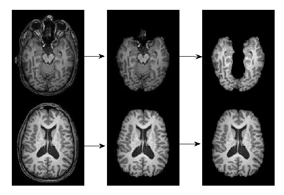
With a set of two-dimensional images, the first step is to remove the skull from the images, then to remove the cerebellum because only cerebrum is needed to calculate. These two steps are shown in Figure 2 from left to right. The right picture contains two different pieces of the MRI brain images. The middle ones are the images with the skull removed, and the right ones are the images with both skull and cerebellum removed.
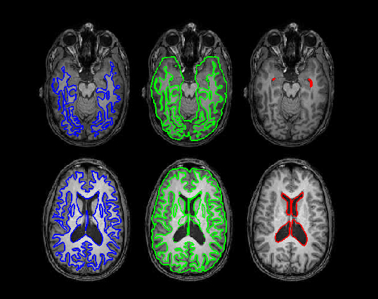
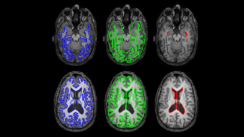
After the pure cerebrum is obtained, an advanced model or algorithm can be applied to those images. Different matters can then be separated, called segmentation. Figure 3 is an example of such a result, where bounded by white contours is the white matter, bounded by the green contour is the gray matter, and bounded by the red contour is the cerebrospinal fluid (CSF).